select language
ACHIEVING HIGH IMAGE QUALITY
  • OVERVIEW
  • IMAGING TECHNOLOGY
  • BENEFITS OF PRODUCTIVITY
  • ADVANCED APPLICATIONS
Call for price
    • Achieving High Image Quality
      Achieving High Image Quality

      High image quality is the most essential factor for MRI systems. Canon (former Toshiba) has developed a range of innovative technologies for Vantage Elan, especially with respect to magnetic field control, which is the key to high image quality.

       
      • The Advanced Magnet system ensures a highly homogeneous static magnetic field
      • Advanced Shielded Gradient Coil generates the most uniform gradient parameters
      • Canon’s unique RF technology transmits high-precision images with minimal noise


    • Comfortable Examinations For All
      Comfortable Examinations For All

      Vantage Elan provides comfort examinations for both operator and patient. M-Power™ helps you navigate effortlessly through each step of the study while Pianissimo™ Σ dramatically reduces acoustic noise to provide patients with a quiet scan.

       
      • Pianissimo Σ enables quiet MRI examinations
      • M-Power guides operators based on clinical workflow, increasing operational efficiency
      • EasyTech provides quick and highly accurate scan positioning
    • Using Your Money Wisely
      Using Your Money Wisely

      The extremely compact system reduces both construction and operating costs which saves your money. Moreover, Eco Mode keeps running costs down and contributes to environmental conservation.

       
      • Requiring only a minimum footprint of 23m2, it is the smallest in its class
      • Rapid installation: completed in as little as 5 days
      • Energy-saving design reduces power requirements by 68%
    • Achieving High Image Quality
      • The Advanced Magnet System Ensures A Highly Homogeneous Static Magnetic Field

      The quality of MR images is dependent on the homogeneity of the static magnetic field generated by the superconducting magnet. Canon (former Toshiba) has developed the Advanced Magnet system, a key technology which generates a highly homogeneous static magnetic field and ensures a wide scanning range and stable image quality.


      • Advanced Shielded Gradient Coil Generates The Most Uniform Gradient Parameters

      The performance of the gradient coil significantly affects image quality. Our Canon Advanced Gradient Shielded Coil System is a completely new concept, which replaces the conventional short gradient coil. Our new gradient coil minimizes eddy currents which results in clearer patient images.


      • High Performance Due To A Micron-Level Cutting Technology

      Canon (former Toshiba) has developed a unique gradient coil cutting technology that cuts high-purity copper ingots into 3D shapes at the micron level. This technology ensures excellent stability and highly effective eddy current suppression.


      • Canon’s Unique RF Technology Transmits High-Precision Images With Minimal Noise

      The RF technology employed in Vantage Elan accurately receives the faint MR signals from the human body, immediately amplifies and then transmits them while suppressing noise, contributing to high image quality.

      These high signals are then converted by direct digitization to make the MRI images.

    • Comfortable Examinations For All
      Comfortable Examinations For All
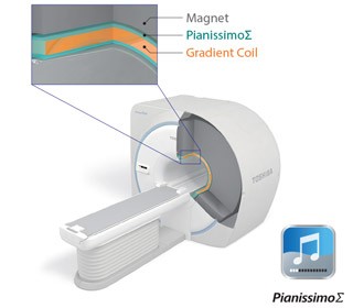
      • Pianissimo Σ Enables Quiet MRI Examinations

      The sound generated during MRI scanning is caused by the vibration of the gradient coil.

      Pianissimo™ Σ reduces the noise level significantly in all types of scanning and provides quiet examinations for patients.


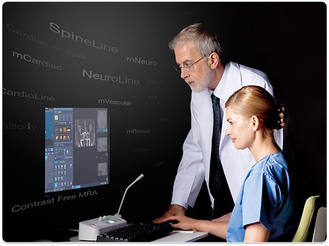
      • M-Power Guides Operators Based On Clinical Workflow, Increasing Operational Efficiency

      With ultimate ease of use, M-Power™ guides user operation according to the workflow, from patient registration to image reconstruction and transfer. A wide variety of applications can support scan positioning and parameter settings, increasing operational efficiency.


      • Directs Facilitates Efficient Examinations Scheduling, Including Patient Registration And Setting Of Scan Conditions

      Orders for MRI examinations can be obtained from the hospital information system using DICOM MWM. The anatomical region to be examined and the protocols to use can be registered in advance.


      • ATLAS Compass Simplifies Routine Examinations

      The system automatically recognizes and selects the coil elements that most efficiently should be used during scanning. This facilitates quicker routine examinations.


      • EasyTech Supports Scan Positioning And Setting Of Scan Conditions

      EasyTech assists scan positioning and condition setting to insure that the optimal positions and conditions will be easily set by any user. Previously parameter settings differed among operators and this resulted in differences in vascular visualization. EasyTech includes DelayTracker, which assists scan condition setting for FBI, and NeuroLine, which assists scan positioning for brain imaging.


      • INSCAN Enables One-Stop Setting Of Scan And Analysis Conditions

      You can set the scan conditions and then the analysis conditions based on the scan result at the same time. Processes from scanning and reconstruction to analysis, which previously required separate steps, can be executed automatically. This streamlines the examination process.


      EasyTEech Provides Quick And Highly Accurate Scan Positioning


      • NeuroLine
        NeuroLine automatically measures and analyzes the shape of the brain, determines the optimal slice position in each plane, and displays them within seconds.


      • SpineLine
        Generally, in spinal examinations, the reference line for the AX plane is manually drawn parallel to the target intervertebral disc or vertebral body. SpineLine automatically measures and analyzes the shape of the spine, determines the positioning ROI in each plane, and displays them within seconds.


    • Using Your Money Wisely
      Using Your Money Wisely
      • Requiring A Minimum Footprint Of Only 23m2, The Vantage Elan Is The Smallest In Its Class

      Not only was the system size reduced, the installation method, cooling method, and control cabinet were innovatively redesigned. Vantage Elan does not require a separate computer room. The overall installation area is approximately 29% smaller than previous 1.5T systems.


      *The minimum footprint may not be applied in all cases depending on each site situation.


      • Rapid Installation: Completed In As Little As 5 Days

      With a new installation, you can start using the system in as little as five days after delivery. Downtime during installation work can be minimized.


      *This installation schedule is a standard schedule for the system and may vary depending on site situation and progress status of the installation.


      • Energy-Saving Design Reduces Power Requirements By 68%

      Total power requirement of Vantage Elan is 25kVA which is the lowest in its class and results in significantly lower running cost. The maximum power consumption is approximately half that of previous models and this also contributes to cost reduction and environmental conservation. In addition, Eco Mode automatically turns off a part of the system when it is not being used. The system can recover from Eco Mode within one second to be ready for scanning.


    • OCTAVE Matrix Coil Concept

      Based on the innovative Atlas matrix coil concept, Canon (former Toshiba) developed an economic version using the similar conceptual matrix design approach. This became the Octave matrix coil concept in which the number of elements was reduced without compromising image quality. For body imaging, the Octave spine coil can be combined with the 4-channel Flex coil or with the Atlas body coil for larger coverage.


      Octave SPEEDER Head/Neck Coil:

      • 11 elements
      • 22cm inner diameter
      • Tiltable with optional wedge


      Octave SPEEDER Spine Coil:

      • Sliding spine coil, integrated in the couch top
      • 12 elements
      • 47cmx 78cm
      • 120cm scanable range for CTL spine when combined with the Octave head coil


    • Orthopedic Multi-Channel SPEEDER Coils

      Shoulder Coil:

      • 6 channels
      • semi-flexible
      • comfortable patient position
      • can also be used for knee, elbow, and ankle


      4-Channel Flex Coil:

      • omni-directional
      • flexible
      • comfortable patient positioning
      • 23cmx 52cm wrap


      Hand/Wrist Coil:

      • 12 elements
      • 6 channels
      • hand and wrist mode
      • ­lateral or isocentric position
      • foldable


    • General Purpose FLEX Coils

      16-Channel Flex Medium Coil:

      • omni-directional
      • flexible
      • comfortable patient positioning
      • 24cmx 40cm wrap


      16-Channel Flex Large Coil:

      • omni-directional
      • flexible
      • comfortable patient positioning
      • 24cmx 70cm wrap


    • MR Mammography

      4-Channel Breast SPEEDER Coil:

      • 4 channels
      • can be equipped with biopsy tools
      • extra large access for biopsies
      • coil elements can be individually positioned


      mBreast Package:

      • Dynamic contrast-enhanced ­examinations
      • diffusion measurements
      • silicone imaging ­combined with the DRKS package also 3D-FFE keyhole imaging
      • time-intensity curves

    • MR Cardio

      mCardiac Package:

      • CINE studies
      • Realtime CINE studies
      • First-pass during rest and stress
      • Late enhancement (2D and 3D)
      • Echo-Navigator coronary angio


      CAAS Package:

      • Cardiac analysis
      • EF, SV, SVI, CO &CI
      • ED & ES volume calculation
      • Body surface area
      • Volume over time
      • Wall motion
      • Cine display
      • Cardiac perfusion
      • Delayed enhancement analysis


close